
How X-ray Scintillators Work, Materials and Applications
How It Works to Use Scintillators for X-ray Imaging
First, what is X-ray Imaging? X-ray imaging is an imaging technique that utilises the penetrating power of X-rays and the differences in X-ray absorption by different materials. It uses a detector to receive transmitted X-rays and convert them into visible images to reveal the internal structure of an object. X-ray, also called X-radiation or known as Roentgen rays, is a type of electromagnetic wave, just like light, only that the X-ray is invisible.
What's making X-ray useful in imaging applications is that X-rays can penetrate substances, X-ray possesses strong penetrating power. X-ray imaging technologies serve a crucial role in fields such as medical treatment, security inspection, industrial flaw detection, and non-destructive testing (NDT).
The degree to which X radiation penetrates substances varies depending on the substance. X-rays can ionise molecules or atoms of the substance, such as the cells of the human body or molecules of the material samples. The interaction between radiation and substances is what makes imaging possible, where we can use detectors to detect signals after passing through the sample being tested and produce an image. The resulting changes in phase, direction, intensities, and wavelength of the radiation reveal information about the internal structure of the object.
Remember, the variable signal detected in the absorption of radiation produces image contrast. For example, in medical X-ray imaging based on a scintillator detector, different tissues in the human body have different X-radiation attenuation coefficients and path lengths within, so that when radiations pass through these human tissues and structures, the radiations are absorbed to varied degrees, resulting in differences in the amount of X-rays reaching the scintillation detector. This creates differences in the amount of light produced from the scintillator across the dimensions of the scintillator, eventually forming images with varied black and white contrast.
X-rays are electromagnetic waves with wavelengths ranging between 0.01~10nm, which implies X-rays are invisible and can scarcely form an image using ordinary photographic substrates. This is where scintillators come in, to be used as X-ray imaging detectors.
Scintillators for X-ray imaging are mainly divided into two types: inorganic scintillators and organic scintillators. Inorganic scintillators are composed of inorganic salt crystals doped with activators, with typical representatives including sodium iodide, cesium iodide, etc. Inorganic scintillators have high density and excellent radiation resistance, and generally high light yield. Organic scintillators are mainly composed of aromatic hydrocarbons with a benzene ring structure, such as anthracene, phenanthrene, and plastic scintillators, etc. Some new materials emit light through aggregation-induced emission (AIE) or thermally activated delayed fluorescence (TADF). Organic scintillators are easy to make into various shapes and can be applied to flexible X-ray imaging.
In scintillator imaging testing, indirect imaging methods are commonly used. In indirect X-ray imaging, X-rays are used as the excitation radiation source and CMOS cameras are used as detectors. We place the sample to be imaged in front of the scintillator, and the X-rays absorbed and transmitted by the sample will be irradiated onto the scintillator. The scintillator converts the X-ray energy into photons and releases them, which are finally captured by the camera for imaging of the sample.
Indirect conversion remains the "time-honoured" solution for mainstream equipment like DR and CT. X-rays are first "absorbed" by scintillator materials, converted into visible light photons, and then captured by an array of photodiodes. However, these photons "diffuse" in all directions within the crystal, gradually diluting spatial resolution; coupled with false signals from dark current, low-contrast details are often "lost in the noise."
Mainstream Scintillator for DR Field: Cesium Iodide (CsI), Gadolinium Oxysulfide (GOS);
CT field: Highlight, GOS, Gemstone, Superlight, etc. names continue to evolve, but the core remains "first convert X-rays into light, then count the light."
Although indirect conversion is relatively simple in principle and somewhat traditional (with issues like light diffusion and noise), it remains the absolute mainstay of X-ray imaging equipment (DR/CT) today due to its mature technology and reliable performance, continuously improved through advancements in materials such as cesium iodide and scintillator detectors.
The working principles of scintillators for X-ray imaging are that scintillators convert X-rays into visible light that can be detected using sensors, and the sensors (such as Si PDs) subsequently convert visible light into electrical signals that can be read out/digitised and form images on the host computer. We can further break down this process of how X-rays are detected using scintillator materials, using GOS:Pr ceramic (praseodymium-doped gadolinium oxysulfide ceramic) as an example:
Here's the physical mechanism of what happens when using X-ray Scintillators. We use GOS:Pr (Pr doped Gadolinium Oxysulfide) scintillator as an example:
1. X-radiation, as a form of ionising radiation enter the scintillator and interacts with Pr:GOS ceramic through physical processes such as the photoelectric effect, Compton scattering, or electron-hole pair generation, depositing energies in the material.
2. The deposited energies lead to the generation of a great deal of fast electrons, which further generate secondary excitations along their tracks. These excitation energies then migrate within the crystal lattice of the GOS:Pr.
3. Energy is ultimately transferred to the luminescent centre.
4. The Pr dopant in GOS:Pr act as an activator that is responsible for the lumination, where energies migrate to the Pr activator ions. The ion in the excited state, after adiative transition, goes back to the ground state and emits fluorescence with a peak emission wavelength of 512 nm.
5. This luminescent signal is converted into an electrical signal using silicon photodiodes (Si PDs) or silicon photomultipliers (SiPMs). The resulting electrical signal can then be processed and analysed to extract information about the incident radiation, thereby enabling detection and imaging.
6. In practical applications, Pr:GOS ceramics are typically fabricated into pixelated arrays. Such structured scintillator arrays facilitate high-resolution imaging in medical diagnostics, security screening, and industrial non-destructive evaluation.
Types of Scintillator Materials for X-ray Imaging
The following table lists the commonly used scintillator materials for X-ray and their technical parameters; the advantages and applications of these scintillators are also evaluated.
Before we start, it is necessary to distinguish between two important concepts in X-ray imaging: hard X-ray and soft X-ray:
-What is hard X-ray imaging and its applications:
Shorter wavelengths than soft X-rays. The wavelength range is typically from 0.01 nanometers to 0.1 nanometers (or 0.1 to 1 angstrom), with high photon energies, generally above 10 keV to 250 keV. It is suitable for deep structural imaging with its excellent penetration. Applications of hard X-ray imaging include CT scans and high-voltage chest radiographs showing the bone and lung structures, astronomical observations, where high absorption efficiencies are required.
-What is soft X-ray Imaging and its applications:
Wavelengths typically range from 0.1 nanometers to 10 nanometers (or 1 to 100 angstroms), with low photon energies, generally between 0.1 keV and 10 keV. Due to their longer wavelengths and lower energy, they have weaker penetrating power and can be easily absorbed by air, skin, and other soft tissues. Soft X-ray imaging has the advantages of being very sensitive to differences in material absorption and good imaging effect on surfaces and thin structures. Soft X-ray imaging are therefore suitable in applications like biological microscopic imaging, semiconductor inspection, and materials science, which call for high spatial resolutions and only require low penetration depth.
Below are the scintillator material types for X-ray imaging. Note that the technical parameters here are only for reference; for more information, please consult Shalom EO's engineers:
| Scintillators for X-ray Imaging | Light Output/Light Yield |
Density(g/cm3) |
Emission Peak Wavelength (nm) |
Decay Time(ns) |
After Glow(%XX@X) |
Advantages and Disadvantages | Applications |
| CsI(Tl) | 52 ph/KeV | 4.51 | 550nm | 1000ns | Afterglow (after 20ms) (%) <0.5 (for normal CsI(Tl)) |
Highest light output, columnar structure guides light, no high voltage required, great match with photodiodes | Most common material for X-ray imaging Not preffered for high-speed applications |
| BGO | 8500 ph/MeV | 7.13 | 480nm | 317ns | NA | A high atomic number indicates a strong stoppeing power, which is beneficial for improving the spatial resolution of detection instruments Low light output |
mainly used in high-energy physics or CT, not for routine low-dose imaging. |
| GOS(Pr) | 27000 ph/MeV | 7.28 | 512nm | 3000ns | Afterglow <0.1%@3ms, ≤0.01%@100ms | Pr doping contributing to speed + low afterglow | High-speed CT Spiral CT and security CT |
| GOS(Tb) | 46500 ph/MeV | 7.34 | 510nm | 60000ns | ≤0.05%@3ms | High luminous efficiencies | Slower than GOS(Pr), suitable for non-high-speed scanning |
| CdWO4 | 12000~15000 ph/MeV |
7.9 | 475nm | 14000ns | Afterglow(%@3ms) <0.1 | High atomic number and high radiation hardness Low afterglow |
Low afterglow desirable cargo scanning, but light output is generally average |
| LaBr3(Ce) | 63000 ph/MeV |
5.2 | 380nm | 25ns | NA | Outstanding energy resolution | limited applications in X-ray imaging, used for nuclide identification |
| YAG(Ce) | 8000 ph/MeV | 4.56 | 530nm | 70ns | NA | Faster decay than CsI Its greatest advantage is that its emission center wavelength is 550nm, which allows for effective coupling with detection devices such as silicon photodiodes |
Can be processed into thin screens for SEM or X-ray microscope |
| GAGG(Ce) | 54000 ph/MeV | 6.63 | 540nm | 90ns | NA | Brightest among oxide microscopes No self-radiation Great match to SiPM Higher price |
Next-generation PET/CT |
What are the Most Important Performance Indicators for X-ray Imaging Scintillators?
For X-ray imaging scintillators, the most important performance indicators are absorption efficiencies, light yield, and spatial resolution (achieved through decay time and afterglow control, and pixelated structure). Meanwhile, the matching degree between the emission spectrum and the sensor determines the system efficiencies, while cost, manufacturability, and environmental stability are key constraints on whether the technology can be applied practically. Future research and development of high-performance scintillators will focus on balancing and optimising these indicators.
Typical Application Scenarios of Scintillator-based X-ray Imaging
1) Medical imaging diagnosis is one of the important application areas of X-ray scintillators, such as CT scanning, X-ray films, etc., all of which rely on scintillators to convert X-rays into visible light and form images.
2) Industrial non-destructive testing (industrial NDT) is also an important application area of X-ray scintillators. By penetrating materials with X-rays and detecting their internal structures, material defects, foreign objects, etc., can be detected. Scintillators play a key role in energy conversion in this process.
3) Security check, especially in baggage screening at airports, stations and other places, X-ray scintillators are widely used in security screening machines to help detect suspicious items, improve security screening efficiency and accuracy.
The Future Development Trend of Scintillators for X-ray Scintillators
New invention of Perovskite nanocrystals:
The development of Perovskite nanocrystals is an important direction of future research in X-ray imaging scintillators. Scientists from the Department of Chemistry, NUS, led by Prof LIU Xiaogang, discovered that lead halide perovskite nanocrystals are highly sensitive to X-ray irradiation. Compared to the traditional scintillator used for X-ray imaging, these Perovskite nanocrystals have much lower growth costs. Another advantage is that the detection limit approx. 400 times lower (only 13 nGyair s-1) than typical doses for medical diagnostics. The material showed great potential for X-ray radiography, where the image can be directly recorded using low-cost, widely accessible digital cameras.
Through innovative materials (such as perovskite nanocrystals), expanded applications (such as therapeutic integration), X-ray imaging scintillators will continue to shine in fields such as medical diagnostics, security inspection, industrial NDT, and even tumour treatment.
High-resolution X-ray imaging via spatially decoupled heavy-atom antennas in organic scintillators:
Organic scintillators, with their advantages such as low cost, sustainabilities and designable structures, hold great promise in the field of X-ray imaging. However, there remains a lack of in-depth understanding regarding the mechanisms of charge transfer and heavy atom design in balancing light yield, decay lifetime, and luminescence bandwidth, which severely limits their practical applications and industrialisation. Professor Tang Benzong's team proposed a spatially separated heavy atom antenna strategy: introducing alkyl bromide units into a localised hybrid charge transfer (HLCT) framework to construct organic scintillators with separated heavy atoms and luminescent centers. This approach maintains efficient X-ray absorption while significantly suppressing non-radiative transitions caused by heavy atoms. Additionally, the molecular system achieves synergistic optimisation of multiple key performance metrics through moderate charge transfer states: short radiative lifetime (3.74 ns), narrow radiative luminescence bandwidth (56 nm), large Stokes shift (110 nm), and 100% photoluminescence quantum yield. Benefiting from these advantages, the scintillator exhibits excellent X-ray luminescence performance, achieving a spatial resolution of 50.0 lp mm-1 in X-ray imaging.
FAQs about X-ray Scintillators:
Q1: What is a scintillator in X-ray imaging?
scintillator in X-ray imaging is a working media that luminesce under radiation of X-ray, people use scintillators to convert invisible X radiations into detectable signals, and utilize the informations carried in these signals to form images that shows details invisible under naked eye, for example, the interiors s of human body (in medical radiography), the inside of baggage (in security check).
Q2: Why are CsI and GOS widely used in X-ray detectors (CsI vs GOS)?
Because these 2 materials have a high Z number (Zeff), high light yield, and an emission spectrum well-matched to sensor.
For the technical parameters of CsI (Tl), GOS(Pr), and GOS(Tb), you can click on the links to check.
In general, CsI have better spatial resolution and light guiding, making it ideal for flat-panel imaging. GOS ceramics are more robust, higher density, easier manufacturing, therefore is the choice for CT and rugged systems.
Q3: What determines the spatial resolution of X-ray scintillators?
Unlike visible light, X-rays are difficult to refract and focus, and can be artificially propagated in a straight line. The spatial resolution of X-ray imaging depends primarily on the relationship between the focal size of the X-ray source, the detector element size, and the distance to the sample.
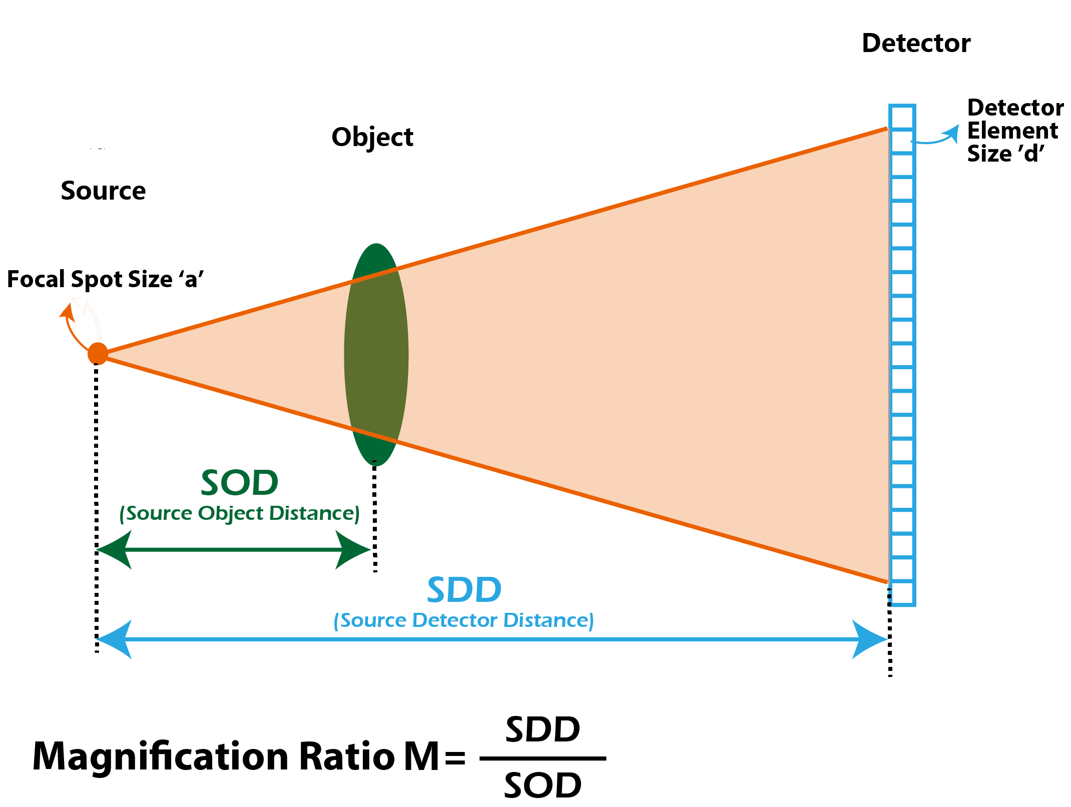
Figure 1. The diagram above shows how an X-ray imaging scintillator are position in application
Q4: What is the difference between soft X-ray and hard X-ray imaging?
Soft X-ray imaging:Uses X-rays with a wavelength range typically from 0.1 nanometers to 10 nanometers (or 10 to 100 angstroms), carrying lower photon energies, generally between 0.1 keV and 10 keV. Due to their lower energy, soft X-rays have low penetrating power and are easily absorbed by matter (even by air, often requiring a vacuum environment). They are primarily used for imaging light elements (like carbon, nitrogen, oxygen) and surface structures, making them ideal for applications in biology (cellular imaging), semiconductor inspection, and material science surface analysis.
Hard X-ray imaging:Uses X-rays with a wavelength range typically from 0.01 nanometers to 0.1 nanometers (or 0.1 to 1 angstrom), carrying higher photon energies, generally above 10 keV to 250 keV (and up to MeV ranges in industrial settings). Due to their high energy, hard X-rays have high penetrating power, allowing them to pass through dense materials like bone, metal, and thick tissues. They are the standard for medical diagnostics (CT scans, radiography), security screening (airport scanners), and industrial non-destructive testing (checking welds or internal defects in castings).
Shalom EO-Supplier of Scintillators for X-ray Imaging
With engineering knowledge and an experienced sales team, Shalom EO offers a comprehensive range of X-ray imaging scintillators for X-ray imaging, including:
· Scintillator materials: CsI, CsI(Tl), CsI(Tl)-Low Afterglow, BGO, LaBr3(Ce), GOS ceramics, CdWO4, etc.
· Pixelated scintillation arrays: made of Tl-doped Cerium Iodide, GAGG(Ce), BGO, CdWO4, GOS(Pr)/GOS(Tb). We are capable of achieving 0.3x0.3mm pixel size, 0.07mm pixel distance

Figure 2. Shalom EO’s pixelated X-ray scintillators
Tags: x ray imaging scintillator